Vittoria Marini, Margalida Campaner Socias, Andreas Dimopoulos, Lorenza Rinvenuto, Enrico Pozzo, Diana Stalkopf, Angelo Serafini, Sara Morri, Ashley Wang, Rita La Rovere, Yoke Chin Chai, Sveva Bollini, Geert Bultynck, H. Llewelyn Roderick, Ionannis Papantoniou, Maurilio Sampaolesi.
Publication: Advanced Healthcare Materials Vol. 15, Issue 16, 24 April 2026
Abstract
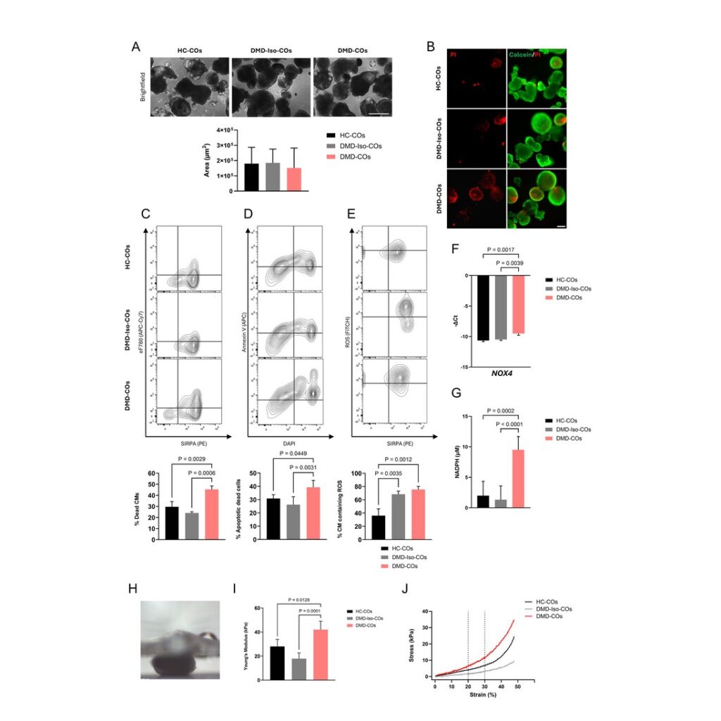
Duchenne muscular dystrophy (DMD) is a genetic disorder characterized by progressive muscle degeneration that significantly reduces the quality of life and lifespan of patients. Currently, cardiomyopathy represents the leading cause of death in later stages of the disease. While calcium dysregulation, fibrosis, and fat deposits are well-documented hallmarks of DMD cardiomyopathies, the exact pathogenic mechanisms remain unclear due to the lack of reliable models. In this study, we developed 3D cardiac organoids (COs) from DMD patient-derived induced pluripotent stem cells (DMD-hiPSCs), their isogenic control (DMD-Iso-hiPSCs), and a healthy hiPSC line (HC-hiPSCs). By day 15 of cardiac differentiation, DMD-COs exhibited key disease features, including increased cell death, elevated ROS levels, and calcium signaling defects, compared with controls. Leveraging bioprinting technology, COs were embedded in a 7% alginate–5% gelatin hydrogel to generate bioprinted constructs (HC-bCOs, DMD-Iso-bCOs, and DMD-bCOs). These constructs self-organized, displaying increased cell–cell communication, and reduced levels of the early cardiac transcription factor NKX2.5. By day 14 post-bioprinting, DMD-bCOs showed increased cell death and dysregulated expression of cardiac and fibrotic markers, mimicking DMD-associated cardiomyopathy. This study demonstrates the potential of both COs and bCOs as a tool for studying DMD cardiomyopathy and advancing drug screening and therapies.
Read the article.